
2023 , Vol. 17 >Issue 06: 340 - 346
DOI: https://doi.org/10.3877/cma.j.issn.1674-0807.2023.06.003
HER-2低表达对乳腺癌新辅助治疗疗效及预后的影响
收稿日期: 2023-02-03
网络出版日期: 2024-01-05
版权
Impact of HER-2 low expression on response of neoadjuvant chemotherapy and prognosis in breast cancer patients
Received date: 2023-02-03
Online published: 2024-01-05
Copyright
探讨HER-2表达状态对乳腺癌新辅助化疗后病理完全缓解(pCR)率以及生存的影响。
根据纳入及排除标准,入组2016年6月1日至2020年12月31日在青岛市市立医院确诊乳腺癌并行新辅助化疗的171例患者进行回顾性研究。根据免疫组织化学(IHC)检测结果,将患者分为HER-2(0)组、HER-2低表达组[IHC染色HER-2(+)或HER-2(2+)且ISH阴性]及HER-2阳性组[IHC染色HER-2(3+)或HER-2(2+)且ISH阳性]。采用χ2检验、Fisher确切概率法或Fisher-Freeman-Halton检验比较3组患者的临床病理特征和预后指标。采用Kaplan-Meier法绘制无瘤生存曲线,用Log-rank检验比较组间生存率。
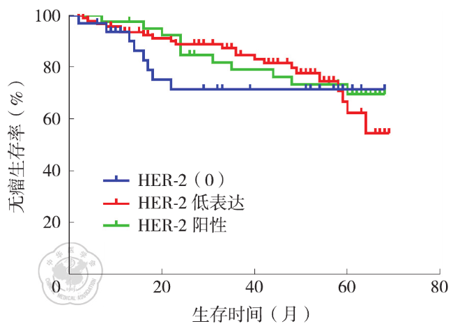
中位随访时间47个月(范围2~54个月)。HER-2(0)组33例(19.3%,33/171),HER-2低表达95例(55.6%,95/171),HER-2阳性43例(25.1%,43/171)。3组患者的激素受体(HR)状态、MP分级比较,差异有统计学意义(χ2=8.497,P=0.014;H=9.406,P=0.009)。新辅助化疗后,HER-2(0)、HER-2低表达、HER-2阳性组的pCR率分别为6.1%(2/33),7.4%(7/95),20.9%(9/43),3组差异无统计学意义(χ2=5.744,P=0.052)。根据HR状态分层,HR阳性患者中,3组的pCR率比较,差异有统计学意义(χ2=7.618,P=0.011),其中HER-2低表达组与HER-2阳性组比较,差异有统计学意义(P=0.033);在HR阴性患者中,3组的pCR率比较,差异无统计学意义(χ2=1.498,P=0.513)。3组的复发转移率分别为24.2%(8/33),23.2%(22/95),25.6%(11/43),差异无统计学意义(χ2=0.097,P=0.953)。HER-2(0)、HER-2低表达、HER-2阳性组的DFS分别为32个月(95%CI:17~57个月)、44个月(95%CI:40~49个月)及58个月(95%CI:56~60个月)。3组的DFS比较,差异无统计学意义(χ2=0.471,P=0.790)。
HER-2低表达不影响乳腺癌患者新辅助化疗后的pCR率,HER-2低表达与HER-2(0)患者的预后比较也没有明显差异。
衣晓丽 , 胡沙沙 , 张彦 . HER-2低表达对乳腺癌新辅助治疗疗效及预后的影响[J]. 中华乳腺病杂志(电子版), 2023 , 17(06) : 340 -346 . DOI: 10.3877/cma.j.issn.1674-0807.2023.06.003
To investigate the effect of HER-2 expression on pathological complete response (pCR) and survival of breast cancer after neoadjuvant chemotherapy.
According to the inclusion and exclusion criteria, 171 patients who were diagnosed with breast cancer and received neoadjuvant chemotherapy in Qingdao Municipal Hospital from June 1, 2016 to December 31, 2020 were enrolled for a retrospective study. According to the immunohistochemical (IHC) results, the patients were divided into HER-2 (0) group, HER-2 low expression group [IHC: HER-2 (+ ) or HER-2 (2+ ) with ISH negative] and HER-2 positive group [IHC: HER-2 (3+ ) or HER-2 (2+ ) with ISH positive]. The clinicopathological features and prognostic parameters were compared among three groups using χ2 test, Fisher exact test or Fisher-Freeman-Halton test. Kaplan-Meier method was used to draw the disease-free survival curve, and log-rank test was used to compare the survival rate between groups.
The patients were followed up for median 47 months (range: 2-54 months). There were 33 cases (19.3%, 33/171) in the HER-2 (0) group, 95 cases (55.6%, 95/171) in HER-2 low expression, and 43 cases (25.1%, 43/171) in HER-2 positive group. The hormone receptor (HR) status and MP grade showed no significant difference among three groups (χ2=8.497, P=0.014; H=9.406, P=0.009). After neoadjuvant chemotherapy, the pCR rates of HER-2 (0), HER-2 low expression and HER-2 positive group were 6.1% (2/33), 7.4% (7/95) and 20.9% (9/43), respectively, indicating no significant difference among three groups (χ2=5.744, P=0.052). According to the stratification of HR status, in HR positive patients, there was a statistically significant difference in pCR rate among three groups (χ2=7.618, P=0.011), and the pCR rate presented a significant difference between HER-2 low expression group and HER-2 positive group (P=0.033). In HR negative patients, there was no significant difference in pCR rate among three groups (χ2=1.498, P=0.513). The recurrence/metastasis rates of the three groups were 24.2% (8/33), 23.2% (22/95), 25.6% (11/43), respectively, with no significant difference (χ2=0.097, P=0.953). The disease-free survival of HER-2 (0), HER-2 low expression and HER-2 positive groups were 32 months (95%CI: 17-57 months), 44 months (95%CI: 40-49 months) and 58 months (95%CI: 56-60 months), respectively. There was no significant difference in disease-free survival among three groups (χ2=0.471, P=0.790).
Low expression of HER-2 cannot affect the pCR rate of breast cancer patients after neoadjuvant chemotherapy, and the patients with HER-2 low expression show no significantly different prognosis compared with HER-2 (0) patients.
表1 不同HER-2表达状态乳腺癌患者的临床病理特征比较 |
| 临床病理特征 | HER-2(0)(33例) | HER-2低表达(95例) | HER-2阳性(43例) | 检验值 | P值 |
|---|---|---|---|---|---|
| 年龄(岁,M (P25,P75)) | 52.0(42.0,60.5) | 50.0(41.0,58.0) | 47.0(39.0,54.0) | H=5.157 | 0.076 |
| 绝经状态[例(%)] | |||||
| 绝经前 | 17(51.5) | 49(51.6) | 28(65.1) | χ2=2.389 | 0.303 |
| 绝经后 | 16(48.5) | 46(48.4) | 15(34.9) | ||
| 病理类型[例(%)] | |||||
| 导管癌 | 27(81.8) | 84(88.4) | 41(95.3) | χ2=5.447 | 0.232 |
| 非导管癌 | 6(18.2) | 11(11.6) | 2(4.7) | ||
| 组织学分级[例(%)]a | |||||
| 1级 | 3(9.1) | 4(4.2) | 1(2.3) | H=5.319 | 0.070 |
| 2级 | 18(54.5) | 53(55.8) | 13(30.3) | ||
| 3级 | 9(27.3) | 29(30.5) | 17(39.5) | ||
| 未知 | 3(9.1) | 9(9.5) | 12(27.9) | ||
| 激素受体[例(%)] | |||||
| 阴性 | 11(33.3) | 21(22.1) | 20(46.5) | χ2=8.497 | 0.014 |
| 阳性 | 22(66.7) | 74(77.9) | 23(53.5) | ||
| Ki-67[例(%)] | |||||
| ≤20% | 10(30.3) | 30(31.6) | 7(16.3) | χ2=3.640 | 0.162 |
| >20% | 23(69.7) | 65(68.4) | 36(83.7) | ||
| ypT[例(%)] | |||||
| T0 | 5(15.2) | 10(10.5) | 9(20.9) | H=0.358 | 0.836 |
| Tis | 0 | 0 | 5(11.6) | ||
| T1 | 19(57.6) | 60(63.2) | 14(32.6) | ||
| T2 | 8(24.2) | 21(22.1) | 10(23.3) | ||
| T3 | 1(3.0) | 4(4.2) | 5(11.6) | ||
| ypN(%) | |||||
| N0 | 12(36.4) | 32(33.7) | 19(44.2) | H=2.328 | 0.312 |
| N1 | 7(21.2) | 25(26.3) | 11(25.6) | ||
| N2 | 10(30.3) | 18(18.9) | 9(20.9) | ||
| N3 | 4(12.1) | 20(21.1) | 4(9.3) | ||
| MP分级[例(%)] | |||||
| 1级 | 3(9.1) | 20(21.1) | 3(7.0) | H=9.406 | 0.009 |
| 2级 | 6(18.2) | 20(21.1) | 9(20.9) | ||
| 3级 | 15(45.4) | 36(37.8) | 12(27.9) | ||
| 4级 | 6(18.2) | 9(9.5) | 5(11.6) | ||
| 5级 | 3(9.1) | 10(10.5) | 14(32.6) | ||
| 化疗方案[例(%)] | |||||
| 蒽环+紫杉类 | 29(87.9) | 83(87.4) | 38(88.4) | χ2=0.028 | 0.986 |
| 紫杉类 | 4(12.1) | 12(12.6) | 5(11.6) | ||
| pCR[例(%)] | |||||
| 是 | 2(6.1) | 7(7.4) | 9(20.9) | χ2=5.744 | 0.052 |
| 否 | 31(93.9) | 88(92.6) | 34(79.1) | ||
| 复发转移[例(%)] | |||||
| 是 | 8(24.2) | 22(23.2) | 11(25.6) | χ2=0.097 | 0.953 |
| 否 | 25(75.8) | 73(76.8) | 32(74.4) |
注:a新辅助化疗后pCR患者的手术标本不再评估组织学分级;pCR为病理完全缓解;MP分级为Miller-Payne分级;对于激素受体状态,HER-2低表达组与HER-2阳性组比较,χ2=8.443, P=0.012;对于MP分级,HER-2低表达组与HER-2阳性组比较,Z=-3.012,P =0.003 |
| [1] |
|
| [2] |
|
| [3] |
|
| [4] |
杨文涛,步宏. 乳腺癌雌、孕激素受体免疫组织化学检测指南[J].中华病理学杂志,2015, 44(4):237-239.
|
| [5] |
《乳腺癌HER2检测指南(2019版)》编写组.乳腺癌HER2检测指南(2019版)[J].中华病理学杂志,2019, 48(3):169-175.
|
| [6] |
中国抗癌协会乳腺癌专业委员会.中国抗癌协会乳腺癌诊治指南与规范(2021年版)[J].中国癌症杂志,2021, 31(10):954-1040.
|
| [7] |
刘裔莎,魏兵,杨雯娟,等.美国癌症联合会乳腺癌分期(第七版)简介[J].中华病理学杂志,2010, 39(11):787-790.
|
| [8] |
|
| [9] |
|
| [10] |
|
| [11] |
|
| [12] |
|
| [13] |
|
| [14] |
|
| [15] |
|
| [16] |
|
| [17] |
|
| [18] |
|
| [19] |
|
| [20] |
|
| [21] |
|
| [22] |
|
| [23] |
|
| [24] |
|
| [25] |
|
| [26] |
|
| [27] |
|
| [28] |
|
| [29] |
于宏,邱芳,顾玺,等. HER-2低表达的三阴性乳腺癌患者的临床特征和预后分析[J]. 中国医科大学学报,2022, 51(8):721-724.
|
/
| 〈 |
|
〉 |