切换至 "中华医学电子期刊资源库"
论著

NF2/YAP信号通路通过FSP1诱导CD24高表达的三阴性乳腺癌细胞铁死亡

  • 蒲卢兰 1 ,
  • 李静佳 2 ,
  • 陈宇 1 ,
  • 周瑜清 1 ,
  • 荣欣欣 1 ,
  • 侯令密 2 ,
  • 周方方 , 1,
展开
  • 1.637000 四川南充,川北医学院基础医学与法医学院
  • 2.637000 四川南充,川北医学院附属医院甲状腺乳腺外科/乳腺癌生物靶向实验室
通信作者:周方方,Email:

收稿日期: 2023-11-14

  网络出版日期: 2024-08-20

基金资助

四川省科技厅自然科学基金面上项目(2022NSFSC0775)

四川省基层卫生事业发展研究中心资助项目(SWFZ22-C-82)

南充市科技局市校科技战略合作项目(20SXQT0052)

川北医学院校级科研发展计划项目(CBY22-QNA11)

版权

未经授权,不得转载、摘编本刊文章,不得使用本刊的版式设计,除非特别声明,本刊刊出的所有文章不代表中华医学会和本刊编委会的观点。本刊为电子期刊,以网刊形式出版。

NF2/YAP signaling pathway induces ferroptosis in triple negative breast cancer cells with high expression of CD24 by FSP1

  • Lulan Pu 1 ,
  • Jingjia Li 2 ,
  • Yu Chen 1 ,
  • Yuqing Zhou 1 ,
  • Xinxin Rong 1 ,
  • Lingmi Hou 2 ,
  • Fangfang Zhou , 1,
Expand
  • 1.School of Basic Medicine and Forensic Medicine, North Sichuan Medical College, Nanchong 637000, China
  • 2.Department of Breast and Thyroid Surgery, Biological Targeting Laboratory of Breast Cancer, Affiliated Hospital of North Sichuan Medical College, Nanchong 637000, China
Corresponding author: Zhou Fangfang, Email:

Received date: 2023-11-14

  Online published: 2024-08-20

Copyright

Copyright by Chinese Medical Association No content published by the journals of Chinese Medical Association may be reproduced or abridged without authorization. Please do not use or copy the layout and design of the journals without permission. All articles published represent the opinions of the authors, and do not reflect the official policy of the Chinese Medical Association or the Editorial Board, unless this is clearly specified.

摘要

目的

研究NF2和YAP表达对CD24高表达的三阴性乳腺癌细胞铁死亡的影响,探讨铁死亡的相关分子机制。

方法

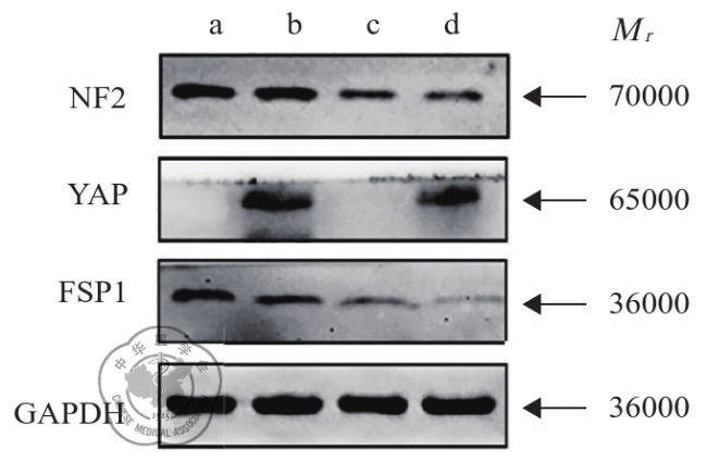
培养CD24high和CD24lowMDA-MB-231细胞,通过台盼蓝染色(TBA)实验检测经半胱氨酸饥饿、铁死亡诱导剂Erastin和RSL3处理后细胞的死亡情况,TBA法检测丙二醛(MDA)含量,分析细胞铁死亡水平。使用Western blot检测NF2和YAP在CD24high和CD24lowMDA-MB-231细胞中的蛋白表达情况,并用慢病毒分别进行敲除和过表达,观察细胞铁死亡水平,并采用实时定量RT-PCR和Western blot检测成纤维细胞特异蛋白1(FSP1)的表达变化。

结果

经半胱氨酸饥饿、铁死亡诱导剂Erastin和RSL3分别处理后MDA-MB-231细胞死亡率增加,CD24low细胞死亡率高于CD24high细胞(t=14.548,P<0.001;t=8.310,P=0.001;t=8.600,P=0.001)。同时,TBA法结果显示,在含或不含半胱氨酸条件下,CD24high和CD24lowMDA-MB-231细胞的MDA含量差异有统计学意义(t=-3.920,P=0.017;t=11.566,P<0.001);与对照组(DMSO)相比,CD24high和CD24low细胞经铁死亡诱导剂Erastin和RSL3处理后MDA水平增加,其中CD24low细胞的MDA含量比CD24high更高(t=10.763,P=0.006;t=24.067,P<0.001)。Western blot实验结果表明,NF2与YAP在CD24low和CD24high细胞的蛋白含量差异有统计学意义(t=-4.331,P=0.012;t=4.219,P=0.013),CD24low细胞中YAP的蛋白表达更高(t=4.219,P=0.013),而CD24high细胞中NF2的蛋白表达更高(t=-4.331,P=0.012)。在CD24highMDA-MB-231细胞中构建NF2敲除或(和)YAP过表达细胞株,并用实时定量RT-PCR和Western blot检测,结果显示铁死亡关键蛋白FSP1在4组间的表达差异有统计学意义(F=30.297,P<0.001)。同时,检测NF2敲除或(和)YAP过表达后CD24highMDA-MB-231细胞的铁死亡情况,结果表明敲除NF2或(和)过表达YAP后加入铁死亡诱导剂Erastin均可在一定程度上促进细胞死亡(F=38.911,P<0.050),其中在NF2敲除联合YAP过表达条件下,CD24highMDA-MB-231细胞死亡率最高,而在Erastin的基础上添加铁死亡抑制剂Fer-1后细胞的死亡率下降,4组间比较差异无统计学意义(F=0.256,P=0.855)。

结论

在CD24高表达的三阴性乳腺癌细胞中敲除NF2联合过表达YAP会抑制FSP1表达,促进细胞铁死亡。

本文引用格式

蒲卢兰 , 李静佳 , 陈宇 , 周瑜清 , 荣欣欣 , 侯令密 , 周方方 . NF2/YAP信号通路通过FSP1诱导CD24高表达的三阴性乳腺癌细胞铁死亡[J]. 中华乳腺病杂志(电子版), 2024 , 18(04) : 206 -211 . DOI: 10.3877/cma.j.issn.1674-0807.2024.04.003

Abstract

Objective

To observe the impact of NF2 and YAP expression on ferroptosis of triple negative breast cancer cells with high expression of CD24 and explore the related molecular mechanism.

Methods

MDA-MB-231 breast cancer cells with high expression of CD24 (CD24high) and low expression of CD24 (CD24low) were cultured. The cells were treated with ferroptosis agonists Erastin, RSL3 and cysteine starvation. The cell death was detected by Trypan blue staining (TBA). Malondialdehyde(MDA)content was measured using the TBA method to analyze the level of cellular ferroptosis. Western blot analysis was used to detect the protein expression of NF2 and YAP in CD24high and CD24low MDA-MB-231 cells. Lentiviral vectors were used to knock out and over-express NF2 and YAP, and changes in cellular ferroptosis were observed. The real-time quantitative RT-PCR and Western blot analysis were used to detect the expression of fibroblast-specific protein 1(FSP1)after NF2 knock out and YAP over-expression.

Results

After treatment with cysteine starvation, the ferroptosis inducers Erastin and RSL3, the death rate of MDA-MB-231 cells increased, with the death rate of CD24low cells significantly higher than that of CD24high cells (t=14.548, P<0.001; t=8.310, P=0.001; t=8.600, P=0.001). Meanwhile, the TBA method results showed that there was a significant difference in MDA content between CD24high and CD24low MDA-MB-231 cells under conditions with or without cysteine (t=-3.920, P=0.017; t=11.566, P<0.001); compared with the control group with DMSO, the MDA levels of CD24high and CD24low cells significantly increased after treatment with the ferroptosis inducers Erastin and RSL3, with the MDA content of CD24low cells significantly higher than that of CD24high cells (t=10.763, P=0.006; t=24.067, P<0.001). Western blot experiments showed that there was a significant difference in the protein content of NF2 and YAP between CD24low and CD24high MDA-MB-231 cells (t=-4.331, P=0.012; t=4.219, P=0.013), with higher expression of YAP protein in CD24low cells (t=4.219, P=0.013) and higher expression of NF2 protein in CD24high cells (t=-4.331, P=0.012). NF2 knockout or (and) YAP overexpression cell lines were constructed in CD24high cells, and real-time quantitative RT-PCR and Western blot analysis were used to detect the expression of the ferroptosis key protein FSP1 among the four groups (F=30.297, P<0.001). Meanwhile, the ferroptosis situation of CD24high MDA-MB-231 cells after NF2 knockout or (and) YAP overexpression was detected, and the results showed that knocking out NF2 or (and) overexpressing YAP could promote cell death to a certain extent after adding the ferroptosis inducer Erastin (F=38.911, P<0.050), with the highest death rate of CD24high MDA-MB-231 cells under the condition of NF2 knockout combined with YAP overexpression. The addition of the ferroptosis inhibitor Fer-1 after Erastin led to a decrease in the death rate of cells, with no statistically significant difference among the four groups (F=0.256, P=0.855).

Conclusions

Knocking out NF2 and overexpressing YAP in CD24high triple negative breast cancer cells can inhibit the expression of FSP1 and promote cell ferroptosis.

三阴性乳腺癌(triple negative breast cancer,TNBC)是侵袭性最高的乳腺癌亚型之一,约占乳腺癌的15%~20%[1],该亚型因缺乏雌激素、孕激素及人表皮生长因子特异性受体,对内分泌治疗及靶向治疗不敏感。目前,化疗是当前大多数TNBC患者的主要治疗措施,但效果差、不良反应大[2,3,4,5]
分化集落因子24(cluster of differentiation,CD24)是一种位于细胞膜的外糖蛋白,与人体多种免疫性疾病相关,具有介导细胞之间、细胞与基质之间黏附的功能。研究发现,CD24与巨噬细胞上的Siglec-10蛋白相互作用传递抑制性信号,能有效逃避巨噬细胞的吞噬[6]。CD24会促进肿瘤细胞的伸展、侵袭,在胃癌、结直肠癌、肝癌等多种恶性肿瘤中呈现不同程度的表达,是未来免疫治疗的潜在靶点[7,8]
铁死亡是一种不同于细胞凋亡、坏死和自噬的程序性细胞死亡[9],它与铁代谢及氧化损伤具有高度相关性,并以胞质和脂质的活性氧明显增多、线粒体体积变小以及膜密度增厚为死亡标志[10]。相关研究表明在肝癌、肾癌、胃癌、卵巢癌、乳腺癌等多种癌细胞中应用铁死亡诱导剂均可有效杀伤肿瘤细胞,抑制肿瘤细胞生长[11,12],提示铁死亡对肿瘤的发展具有一定的抑制作用,可作为肿瘤治疗的新方法。由于肿瘤的耐药性会对铁死亡产生抵抗力,通过抑制纤维细胞特异蛋白1(fibroblast specific protein 1, FSP1)、谷胱甘肽过氧化物酶4(glutathione peroxidase 4, GPX4)、N-脂肽甲基转移酶(N-myristoyltransferase, NMT)或者其他促进铁死亡的治疗方法可增强抑制效果[13,14,15]。研究报道实体瘤内部肿瘤细胞中特异性敲除NF2或过表达YAP,可有效提升肿瘤细胞对铁死亡的敏感性[16,17]。目前铁死亡在高表达CD24(CD24high)的MDA-MB-231细胞中的研究较少,因此,本研究通过调节NF2-YAP信号通路,观察其对CD24highMDA-MB-231细胞铁死亡的影响,探索这类细胞的主要死亡方式,以期为临床TNBC患者提供新的治疗方案。

材料与方法

一、细胞与主要试剂

CD24low和CD24highMDA-MB-231细胞由课题组前期研究留存[5];DMEM培养基和血清购自美国Gibco公司;NF2敲除和YAP过表达质粒由南京金斯瑞生物技术有限公司构建;RNA逆转录试剂购自美国Themo公司;TB-green荧光定量试剂盒购自日本Takara公司;实验所需引物由上海生工合成;BCA蛋白浓度测定试剂盒和丙二醛(malondialdehyde,MDA)检测试剂盒购自北京碧云天生物技术公司;抗NF2抗体、YAP抗体和FSP1抗体构自美国Cell Signaling Technology公司;抗GAPDH抗体购自武汉Proteintech公司;铁死亡抑制剂1(ferrostatin-1,Fer-1)、RSL3和Erastin试剂购自美国MedChemExpress公司。其他所需试剂或耗材为本实验室配制或配备。

二、细胞系培养与构建

CD24low和CD24highMDA-MB-231细胞,采用含10%胎牛血清的DMEM培养基(添加1%青霉素/硫酸链霉素)于37 ℃、5% CO2恒温培养箱内培养。NF2敲除和YAP过表达细胞系均使用CRISPR/Cas9方法构建,通过Delta-VPR和CMV-VSV-G慢病毒系统包装质粒转染293T细胞。12 h后更换培养基,48 h后收集上清液并通过0.45 μm过滤器去除细胞。然后将细胞与滤液共培养过夜(添加4 μg/ml polybrene),并换为完全培养基。48 h后,观察荧光转染效率,继续培养并提取RNA和蛋白质以便后续实验。NF2单链引导RNA(single guide RNA, sgRNA)序列:CCTGGCTTCTTACGCCGTCC;YAP sgRNA序列:ACTTCTATGCTGAATCAAGT。

三、Western blot

使用10%~15% SDS-PAGE凝胶分离蛋白质并转移到PVDF膜上,然后用5%脱脂乳在37℃下封闭1 h,4℃分别孵育一抗NF2(1∶1000)、YAP(1∶1000)、FSP1(1∶1000)、GAPDH(1∶1000)过夜,用TBST洗涤3次,每次10 min;二抗在37 ℃下孵育2 h,用TBST洗涤3次,每次10 min,随后进行化学发光检测,实验重复3次。

四、实时定量RT-PCR

收集细胞,提取总RNA,按照逆转录试剂说明书将RNA逆转录为cDNA,进一步按照TB Green® Premix Ex TaqTMⅡ(Tli RNaseH Plus)试剂盒配制实时定量RT-PCR反应体系,总反应体系为20 μl。反应结束后,通过溶解曲线的Tm值来确定PCR产物的特异性,采用2-△△CT法进行分析,得出目标基因的转录水平,实验重复3次。引物序列情况见表1
表1 实时荧光定量反转录PCR引物序列
引物名称 引物序列(5’→3’)
NF2 正向:GTGCCATACTAAGCAGCTTGC
反向:ACACTACAGCTCGGAAATCCA
YAP 正向:GTCGGAACCCTTCCCTCATTA
反向:GGCCTTCACTGACAGGTAGTA
FSP1 正向:ATCGTCCGAGATGATGTTGATGGG
反向:ATTACTGCTACTCCGTACACCACTG
GAPDH 正向:TGACTTCAACAGCGACACCCA
反向:CACCCTGTTGCTGTAGCCAAA

五、铁死亡的诱导和抑制

通过半胱氨酸饥饿、Erastin或RSL3诱导细胞铁死亡。半胱氨酸饥饿诱导方法为:用PBS洗涤细胞3次,添加不含半胱氨酸的培养基(10% FBS)并培养30 h。Erastin或RSL3诱导方法为直接向培养基中添加10 μm Erastin或5 μm RSL3处理24 h。铁死亡的抑制方法为在添加铁死亡诱导剂22 h后,再添加铁死亡抑制剂Fer-1处理2 h。实验重复3次。

六、细胞死亡和活力检测

细胞死亡和活力情况通过台盼蓝染色实验检测。在未经任何处理的CD24high细胞内加入DMSO(1∶1000)作为对照组,以NF2敲除(NF2-KO)、YAP过表达(YAP-OV)和NF2敲除联合YAP过表达(YAP-OV+NF2-KO)细胞组作为实验组。选择目的细胞制备细胞悬液,将细胞悬液与0.4%台盼蓝溶液以9∶1混合,染色3 min,显微镜下观察死细胞比例并计数。活细胞率(%)=活细胞总数/(活细胞总数+死细胞总数)×100%。实验重复3次。

七、细胞MDA水平检测

将细胞接种于96孔板上,每组设置6个复孔,处理后按照MDA试剂盒操作说明书检测细胞内MDA水平,用酶标仪检测532 nm波长下的吸光度值,计算MDA含量,实验重复3次。

八、统计学分析

结果采用SPSS 26.0统计软件进行分析,部分实验数据采用GraphPad软件绘制统计图。计量资料用±s表示,独立样本或组间比较采用t检验,多组间比较采用单因素方差分析(两两比较采用LSD校正)。双侧检验,以P<0.050为差异有统计学意义。

结果

一、铁死亡诱导剂可以提高MDA-MB-231细胞的死亡率

CD24low和CD24highMDA-MB-231细胞分别经半胱氨酸饥饿、Erastin和RSL3诱导后,细胞的死亡率增加。结果表明,在含半胱氨酸条件下培养CD24high和CD24low细胞,细胞死亡率差异无统计学意义(t=1.301,P=0.263),经半胱氨酸饥饿处理后细胞死亡率增加,CD24low细胞死亡率比CD24high细胞更高(t=14.548,P<0.001),具体数据见表2。以DMSO为对照培养CD24high和CD24low细胞,细胞死亡率差异无统计学意义(t=1.292,P=0.266),经铁死亡诱导剂Erastin处理后细胞死亡率增加,CD24low细胞死亡率比CD24high细胞更高(t=8.310,P=0.001),经铁死亡诱导剂RSL3处理后细胞死亡率增加,CD24low细胞死亡率比CD24high细胞死亡率更高(t=8.600,P=0.001),具体数据见表3
表2 半胱氨酸饥饿处理后CD24low与CD24high细胞死亡率比较
组别 实验次数 含半胱氨酸 半胱氨酸饥饿
CD24low 3 4.60±0.63 28.93±0.40
CD24high 3 3.83±0.81 12.60±1.82
t   1.301 14.548
P   0.263 <0.001
表3 铁死亡诱导剂处理CD24low与CD24high细胞死亡率比较
组别 实验次数 DMSO Erastin RSL3
CD24low 3 4.93±1.36 38.15±2.92 39.51±2.16
CD24high 3 3.61±1.12 14.73±3.91 18.90±1.79
t   1.292 8.310 8.600
P   0.266 0.001 0.001

二、铁死亡诱导剂可增加MDA-MB-231细胞的MDA水平

半胱氨酸和铁死亡诱导剂会影响MDA-MB-231细胞的MDA水平。在半胱氨酸条件下培养CD24high和CD24low细胞,CD24high细胞MDA含量比CD24low细胞更高(t=-3.920,P=0.017),但经半胱氨酸饥饿处理后,CD24low细胞MDA含量比CD24high细胞更高(t=11.566,P<0.001),具体数据见表4。后续实验进一步使用铁死亡诱导剂处理CD24high和CD24low细胞,以DMSO为对照培养CD24high和CD24low细胞,细胞死亡率差异无统计学意义(t=0.970,P=0.387),经铁死亡诱导剂Erastin处理后细胞MDA水平增加,CD24low细胞MDA含量比CD24high细胞更高(t=10.763,P=0.006),经铁死亡诱导剂RSL3处理后细胞MDA水平增加,CD24low细胞MDA含量比CD24high细胞更高(t=24.067,P<0.001),具体数据见表5
表4 半胱氨酸饥饿处理后CD24low与CD24high细胞MDA水平比较
组别 实验次数 含半胱氨酸 半胱氨酸饥饿
CD24low 3 0.32±0.02 1.63±0.06
CD24high 3 0.37±0.01 0.96±0.08
t   -3.920 11.566
P   0.017 <0.001
表5 铁死亡诱导剂处理后CD24low与CD24high细胞MDA水平比较
组别 实验次数 DMSO Erastin RSL3
CD24low 3 0.42±0.04 1.76±0.02 1.67±0.02
CD24high 3 0.40±0.03 1.28±0.08 1.22±0.03
t   0.970 10.763 24.067
P   0.387 0.006 <0.001

三、CD24highMDA-MB-231细胞中NF2高表达及YAP低表达

Western blot检测CD24low与CD24high细胞中NF2和YAP蛋白水平。结果表明,CD24low细胞中NF2的蛋白水平明显低于CD24high细胞(6.97±4.08比19.08±2.61,t=-4.331,P=0.012),YAP的蛋白水平明显高于CD24high细胞(28.57±3.98比18.31±1.38,t=4.219,P=0.013)(图1)。
图1 Western blot检测CD24low和CD24highMDA-MB-231细胞中NF2和YAP蛋白表达情况

四、NF2敲除和YAP过表达能够抑制CD24high MDA-MB-231细胞铁死亡关键蛋白FSP1的表达

CD24highMDA-MB-231细胞铁死亡关键抑制蛋白FSP1表达水平受NF2和YAP的影响。实时定量RT-PCR结果显示,CD24high细胞中FSP1的mRNA表达量为15.38±2.12,经YAP过表达或(和)NF2敲除处理后细胞中FSP1的mRNA表达降低(9.83±0.11,10.87±0.89,6.24±0.55)。单因素方差分析结果显示,FSP1的mRNA表达在4组间比较具有统计学意义(F=30.297,P<0.001);两两比较(LSD校正)结果显示,对照组与YAP-OV组(P<0.001)、NF2-KO组(P=0.002)、YAP-OV+NF2-KO(P<0.001)组之间差异具有统计学意义,YAP-OV组与NF2-KO组之间差异不具有统计学意义(P=0.315),与YAP-OV+NF2-KO组之间差异有统计学意义(P=0.006),NF2-KO组与YAP-OV+NF2-KO组之间差异具有统计学意义(P=0.001)。同时,Western blot检测结果也进一步表明,FSP1在单独NF2敲除或YAP过表达后的蛋白水平降低,在NF2敲除联合YAP过表达后下降更为明显(图2)。
图2 NF2敲除联合YAP过表达降低CD24highMDA-MB-231细胞中FSP1表达

注:a代表对照组;b代表YAP过表达组(YAP-OV);c代表NF2敲除组(NF2-KO);d代表NF2敲除联合YAP过表达组(YAP-OV+NF2-KO)

五、NF2敲除联合YAP过表达促进CD24highMDA-MB-231细胞铁死亡

对照组、NF2-KO组、YAP-OV组和YAP-OV+NF2-KO组的细胞死亡率,4组间比较,差异无统计学意义(F=0.469,P=0.712);经铁死亡诱导剂Erastin处理后各组细胞死亡率均有不同程度增加,且4组间比较差异具有统计学意义(F=38.911,P<0.001);两两比较(LSD校正),其中对照组与YAP-OV组(P=0.005)、NF2-KO组(P<0.001)、YAP-OV+NF2-KO组(P<0.001)之间差异具有统计学意义,YAP-OV组与NF2-KO组(P=0.026)、YAP-OV+NF2-KO组(P<0.001)之间差异具有统计学意义,NF2-KO组与YAP-OV+NF2-KO组之间差异具有统计学意义(P=0.004)。铁死亡诱导剂Erastin的基础上添加铁死亡抑制剂Fer-1后细胞的死亡率下降,4组间比较差异无统计学意义(F=0.256,P=0.855),具体数据见表6
表6 NF敲除联合YAP过表达CD24high细胞加药处理后死亡率比较
组别 实验次数 DMSO Erastin Erastin+Fer-1
对照 3 5.74±0.50 19.89±0.68 6.37±2.50
YAP-OV 3 6.64±1.33 29.43±0.53 6.68±0.78
NF2-KO 3 5.86±1.34 36.31±6.10 7.42±1.48
YAP-OV+NF2-KO 3 6.19±0.60 46.28±0.57 7.01±0.71
F   0.469 38.911 0.256
P   0.712 <0.001 0.855

讨论

乳腺癌是女性常见的恶性肿瘤,病理类型多样。TNBC是指ER、PR和HER-2表达均阴性的乳腺癌,好发于小于40岁的年轻女性,约占所有乳腺癌的10%~22%,其侵袭性、复发率和死亡率均高于其他类型的乳腺癌,且预后较差[1,2,3,17,18]。CD24是一种低分子量高度糖化的蛋白质,与肿瘤细胞增殖、生长、凋亡和转移密切相关,在卵巢肿瘤、乳腺肿瘤和B细胞淋巴瘤等多种肿瘤中高表达[19,20,21]。笔者前期实验结果显示乳腺癌患者中CD24高表达,且又以在TNBC中CD24特异性高表达最为明显[7]。铁死亡是一种区别于传统的细胞凋亡、坏死及细胞自噬的细胞死亡方式。相关研究表明铁死亡在乳腺癌中起着重要作用,通过激活铁死亡途径能抑制乳腺癌细胞增殖,改善其对化疗药物的耐药性,增强对放射治疗的敏感性及抑制癌细胞远处转移,可作为乳腺癌患者治疗的潜在新靶点[15]。然而,有关CD24高表达TNBC细胞的铁死亡调控机制尚不清楚。
本研究通过检测CD24high和CD24lowMDA-MB-231细胞在不同铁死亡诱导剂作用下,细胞的死亡率、存活率和MDA含量来反应铁死亡水平。研究提示,铁死亡是杀伤CD24high细胞的有效途径。Wu[17]等的研究表明NF2-YAP信号通路对肿瘤细胞铁死亡起重要调节作用,特异性敲除NF2或过表达YAP,可有效提升肿瘤对铁死亡的敏感性。本实验结果显示与CD24low细胞相比,CD24high细胞中NF2高表达,而与之对应的YAP则低表达,且CD24high细胞死亡方式以铁死亡为主。因此,笔者推测NF2-YAP信号通路在CD24highMDA-MB-231细胞铁死亡中具有重要调节作用,并进行系列实验分析NF2-YAP信号通路在CD24highMDA-MB-231细胞铁死亡中的作用。
FSP1作为一种铁死亡关键抑制蛋白,可通过抑制铁离子的积累和减少氧化应激反应来保护细胞免受铁死亡的损伤,阻止细胞发生铁死亡[13]。Doll等[22]研究发现,FSP1高表达与铁代谢紊乱和铁死亡的增加相关,可能促进肿瘤的生长和进展。因此,FSP1被认作一种肿瘤铁死亡抑制因子,通过抑制FSP1的功能,可以增加肿瘤细胞的铁死亡敏感性。之前的研究表明,铁死亡可以通过钙黏蛋白介导的细胞互作进行自主调节,这一过程受NF2/YAP信号通路调控[17]。针对NF2/YAP通路对FSP1的调控作用,研究发现,NF2蛋白在与YAP结合后,会促使YAP从细胞核转移到细胞质,从而抑制YAP的转录活性[23]。YAP作为一种转录共激动剂可与多种DNA结合转录因子结合,若抑制YAP的转录活性会改变特定基因的转录活性[24,25]。此外,NF2/YAP信号通路可以与Wnt/β-catenin信号通路相互作用,而FSP1的表达受该信号通路的调控[26,27]。在本研究中,经NF2敲除联合YAP过表达处理后,Erastin对CD24high细胞的杀伤效果增强,同时FSP1的表达减弱,提示NF2/YAP通路可能通过抑制FSP1的表达促进CD24high细胞铁死亡。
铁死亡作为一种新发现的细胞死亡形式,在杀死多种肿瘤细胞中发挥重要作用,是目前肿瘤研究的热点之一[28]。乳腺癌的病因和有效治疗手段目前尚未完全弄清,而本研究发现,NF2/YAP信号通路可通过抑制FSP1表达来激活铁死亡途径,从而有效杀伤CD24high细胞,该信号通路可能是调控CD24highMDA-MB-231细胞铁死亡的关键信号通路。这为TNBC的治疗提供了新思路和潜在靶点,但具体的调控机制仍需要实验验证。
[1]
Franzoi MA, Romano E, Piccart M. Immunotherapy for early breast cancer: too soon, too superficial, or just right?[J].Ann Oncol, 2021, 32(3):323-336.

[2]
Waks AG, Winer EP. Breast cancer treatment[J]. JAMA, 2019, 321(3):316.

[3]
Stockwell BR, Jiang X, Gu W. Emerging mechanisms and disease relevance of ferroptosis[J]. Trends Cel Biol, 2020, 30(6):478-490.

[4]
Mou Y, Wang J, Wu J, et al. Ferroptosis, a new form of cell death: opportunities and challenges in cancer[J]. J Hematol Oncol, 2019, 12(1):34.

[5]
Hou L, Pu L, Chen Y, et al. Targeted intervention of NF2-YAP signaling axis in CD24-overexpressing cells contributes to encouraging therapeutic effects in TNBC[J]. ACS Nano, 2022, 16(4):5807-5819.

[6]
Barkal AA, Brewer R, Markovic M, et al. CD24 signalling through macrophage siglec-10 is a target for cancer immunotherapy [J]. Nature, 2019, 572(7769):392-396.

[7]
Pu L, Wang D, Zhou Y, et al. Expression of CD24 in triple negative breast cancer and its relationship with EMT[J]. Modern Oncol, 2023, 31(2):217-221.

[8]
Li L, Gong Y, Tang J, et al. ZBTB28 inhibits breast cancer by activating IFNAR and dual blocking CD24 and CD47 to enhance macrophages phagocytosis[J]. Cell Mol Life Sci, 2022, 79(2):83.

[9]
Stockwell BR, Jiang X, Gu W. Emerging mechanisms and disease relevance of ferroptosiss[J]. Trends Cell Biol, 2020, 30(6):478-490.

[10]
Guo Y, Chen G. Research progress on iron death regulatory mechanism and tumor occurrence and treatmens[J]. Adv Physiol Sci, 2019, 50(2):88-93.

[11]
Liang C, Zhang X, Yang M, et al. Recent progress in ferroptosis inducers for cancer therapys[J]. Adv Mater, 2019, 31(51):e1904197.

[12]
Xie X, Fan T, Zhang B, et al. Research progress on the correlation between iron death and breast cancer treatments[J]. Chin J Clin Oncol, 2020, 47(8):423-426.

[13]
Doll S, Freitas FP, Shah R, et al. FSP1 is a glutathione-independent ferroptosis suppressors[J]. Nature, 2019, 575(7784):693-698.

[14]
Bersuker K, Hendricks JM, Li Z, et al. The CoQ oxidoreductase FSP1 acts parallel to GPX4 to inhibit ferroptosiss[J]. Nature, 2019, 575(7784):688-692.

[15]
Kosciuk T, Lin H. N-Myristoyltransferase as a glycine and lysine myristoyltransferase in cancer, immunity, and infectionss[J]. ACS Chem Biol, 2020, 15(7):1747-1758.

[16]
Xu F, Chen WD, Guo MH, et al. Progress of ferroptosis mechanism and application in lung cancer therapys[J]. J Basic clinMed, 2021, 41:442.

[17]
Wu J, Minikes AM, Gao M, et al. Intercellular interaction dictates cancer cell ferroptosis via NF2-YAP signallings[J]. Nature, 2019, 572(7769):402-406.

[18]
Coates JT, Sun S, Leshchiner I, et al. Parallel genomic alterations of antigen and payload targets mediate polyclonal acquired clinical resistance to sacituzumab govitecan in triple-negative breast cances[J]. Cancer Discov, 2021, 11(10):2436-2445.

[19]
Gilad N, Zukerman H, Pick M, et al. The role of CD24 in multiple myeloma tumorigenicity and effects of the microenvironment on its expressions[J]. Oncotarget, 2019, 10(52):5480-5491.

[20]
Ni YH, Zhao X, Wang W. CD24, a review of its role in tumor diagnosis, progression and therapys[J]. Curr Gene Ther, 2020, 20(2):109-126.

[21]
Dong F. Meta-analysis of the expression of CD24 in breast cancer and its correlation with clinical pathological characteristics and prognosis[D]. Jilin University, 2020, 1-35.

[22]
Doll S, Freitas FP, Shah R, et al. FSP1 is a glutathione-independent ferroptosis suppressors[J]. Nature, 2019, 575(7784):693-698.

[23]
Meng Z, Moroishi T, Guan KL. Mechanisms of hippo pathway regulations[J]. Genes Dev, 2016, 30(1):1-17.

[24]
Huang LS, Sudhadevi T, Fu P, et al. Sphingosine kinase 1/S1P signaling contributes to pulmonary fibrosis by activating Hippo/YAP pathway and mitochondrial reactive oxygen species in lung fibroblastss[J]. Int J Mol Sci, 2020, 21(6):2064.

[25]
Liu-Chittenden Y, Huang B, Shim JS, et al. Genetic and pharmacological disruption of the TEAD-YAP complex suppresses the oncogenic activity of YAPs[J]. Genes Dev, 2012, 26(12):1300-1305.

[26]
Zhang R, Gao Y, Zhao X, et al. FSP1-positive fibroblasts are adipogenic niche and regulate adipose homeostasiss[J]. PLoS Biol, 2018, 16(8):e2001493.

[27]
Wang D, Dai C, Li Y, et al. Canonical Wnt/β-catenin signaling mediates transforming growth factor-β1-driven podocyte injury and proteinurias[J]. Kidney Int, 2011, 80(11):1159-1169.

[28]
Mou Y, Wang J, Wu J, et al. Ferroptosis, a new form of cell death: opportunities and challenges in cancers[J]. J Hematol Oncol, 2019, 12(1):34.

文章导航

/


AI


AI小编
你好!我是《中华医学电子期刊资源库》AI小编,有什么可以帮您的吗?